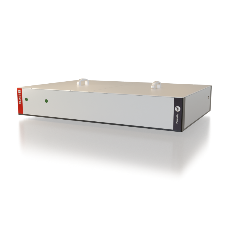
White Dwarf dual
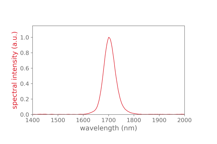
- wavelength: 1300 nm and 1700 nm
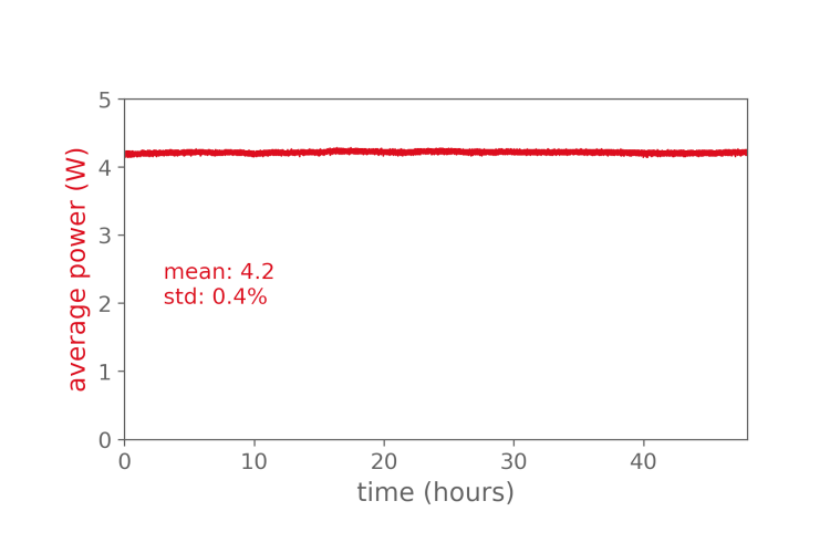
- average power: > 3 W or > 5 W
- optimized for multicolor deep imaging
- based on industrial Yb-fiber femtosecond laser
Introduction
High performance femtosecond laser for three-photon microscopy (3p microscopy)
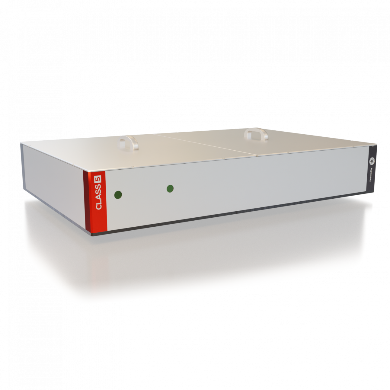
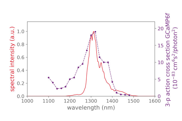
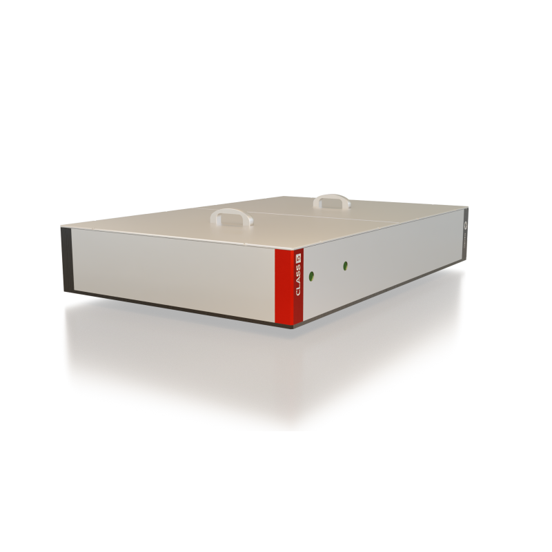
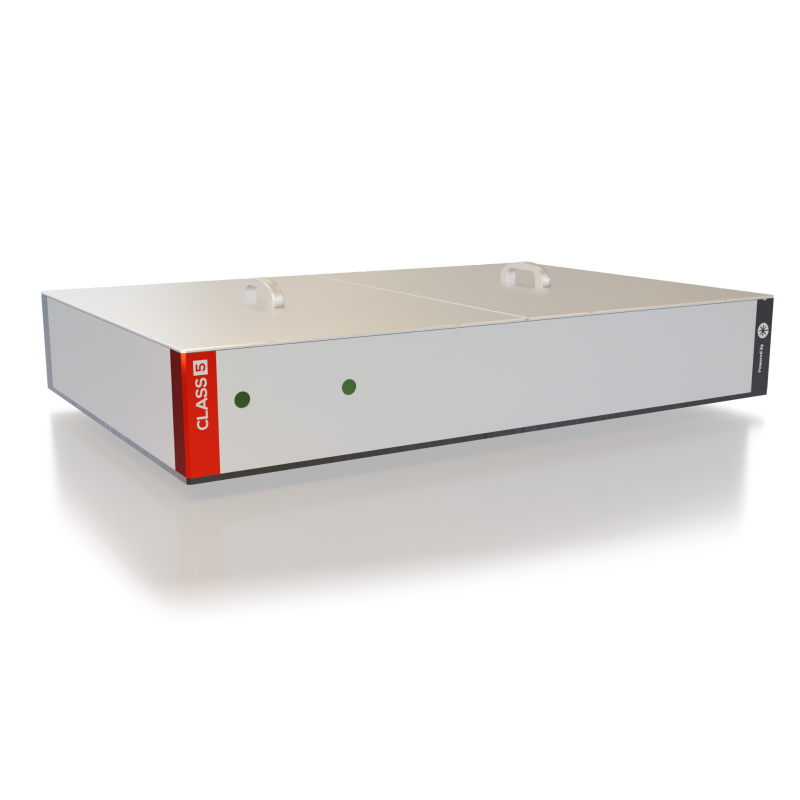
The White Dwarf is a compact and robust femtosecond laser system specially designed for 3-photon microscopy, offering now simultaneously 1300 and 1700 nm at outstanding performance parameters. With the highest peak power available for 3-photon microscopy, the White Dwarf allows neuroscientists to image deeper, faster and at better resolution than ever before at green and red fluorescent markers.

By loading the video, you agree to YouTube’s privacy policy.
Learn more
Features
Optimized for multi-color imaging
Ideal for deep imaging
Proven in the field
Reliability
1035 nm output
Specs
| WD-1300-1700 | WD-1300-1700-pro | |
|---|---|---|
| Central wavelength | 1300 nm and 1700 nm (switchable or simultaneous) | 1300 nm and 1700 nm (switchable or simultaneous) |
| Average power | > 3 W | > 5 W |
| Pulse duration FWHM |
< 50 fs at 1300 nm and < 65 fs at 1700 nm | < 50 fs at 1300 nm and < 65 fs at 1700 nm |
| Dispersion compensation optional |
up to -12.000 fs^2 at 1300 nm and up to +/-4.000 fs^2 at 1700 nm | up to -12.000 fs^2 at 1300 nm and up to +/-4.000 fs^2 at 1700 nm |
| Pulse energy compressed |
> 3 µJ at 1 MHz | > 5 µJ at 1 MHz |
| Repetition rate | up to 2 MHz | up to 2 MHz |
| Beam quality M2 | < 1.3 | < 1.3 |
| Power stability 12 hrs |
< 1 % | < 1 % |
| Dimensions | 80 cm x 120 cm | 80 cm x 120 cm |
| Pump laser incl. | Coherent Monaco 40 W | Coherent Monaco 60 W |
Performance
Applications
-
Volumetric brain imaging
-
Multi-color imaging
-
Adaptive optics (AO) three-photon microscopy
FAQ
Yes, the White Dwarf is already installed and tested by many labs. Please contact us, we are happy to share specific references to your application. You can find a first overview of scientific publications from our users below.
The White Dwarf dual can offer up to three outputs: 1300 nm, 1700 nm and 1035 nm. The system can be designed to offer all outputs simultaneously or switchable between different outputs. Please contact us for customization options.
Our White Dwarf dual version can be configured at any single repetition rate between 1 and 2 MHz. The maximum average power is available at the configured repetition rate. Lower repetition rates can be chosen using the built-in pulse-picker. This provides a constant pulse energy at correspondingly lower average power. Example: WhiteDwarf operating at 2 MHz and 2.5 µJ delivers 5 W; it can be down-picked to 1 MHz and 2.5 µJ delivering 2.5 W.
Our service team will install the pump laser and the OPCPA at the customer’s site. Users will receive training on the operation of the White Dwarf.
Yes, we offer the full package including pump laser and OPCPA. The pump laser is integrated into the OPCPA housing on a shared thermally managed breadboard offering a robust one-box solution.
Publications
-
May 2, 2019Weisenburger et al., Volumetric Ca2+ Imaging in the Mouse Brain Using Hybrid Multiplexed Sculpted Light Microscopy, Cell (2019)
-
September 30, 2021Streich, L., Boffi, J.C., Wang, L. et al. High-resolution structural and functional deep brain imaging using adaptive optics three-photon microscopy. Nat Methods 18, 1253–1258 (2021).
-
December 21, 2023Amr Tamimi, Martin Caldarola,…, Robert Prevedel, Deep mouse brain two-photon near-infrared fluorescence imaging using a superconducting nanowire single-photon detector array
