Volumetric imaging of deep brain tissue
One of the main goals in neuroscience is to capture functional dynamics of in-vivo neuronal circuits in deep brain tissue.
In particular, multi-photon microscopy reduces the out-of-focus excitation within tissue and shows a good signal-to-background ratio at deep sample depths, because of higher order nonlinear excitation and weaker scattering at longer wavelengths.
The high average power of the White Dwarf OPCPA powered by Coherent allows to discover the full potential of 3D bio-imaging in a large volume while maintaining single-cell resolution.
Volumetric imaging methods benefit from the synchronized dual output option at 960 nm and 1300 nm to record simultaneously two- and three-photon excitations of green fluorescent proteins. The high pulse energy available allows spatiotemporal multiplexing and recording of neuroactivity beyond one milimeter deep within mouse brain.
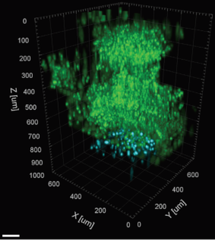
Additionally, the White Dwarf OPCPA can be configured for three-photon excitation of red fluorescent proteins at 1700 nm. Pulse energies of more than 2 µJ at 1 MHz repetition rate allow fast imaging of deep-tissue.
The White Dwarf OPCPA implements the Coherent Monaco industrial fiber laser as pump source for long-term stability, reliability and turn-key operation.


